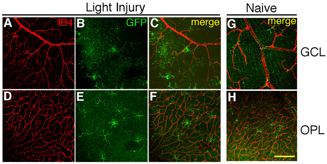
Fig. 8.
Light injury causes a change in the distribution and number of GFP+ cells. (A–C) Optical sections from the GCL. (D–F) Same image field as in A–C, but the optical sections were taken from the OPL. (A, D) Blood vessels stained with IB4 (red). (B, E) GFP+ cells (green). (C, F) Merged. (G) GFP+ cells in the GCL of naive retinas. (H) OPL from the same field as G. Scale bar, 50 µm. Cell counts are shown in Table 3.

