
Fig. 4.
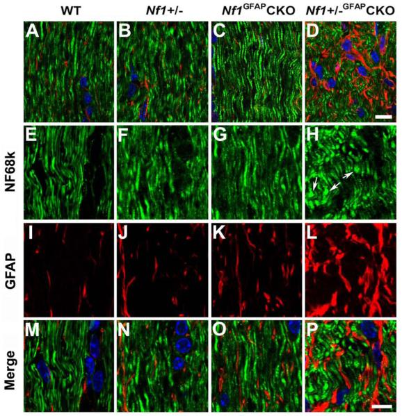
Abnormal axon architecture in Nf1+/−GFAPCKO mice. Sections from 9-month-old wild-type (A, E, I, M), Nf1+/− (B, F, J, N), Nf1GFAPCKO (C, G, K, O) and Nf1+/−GFAPCKO (D, H, L, P) mice were double labeled with Neurofilament 68k and GFAP antibodies. Well-oriented axonal fiber projections and GFAP-positive astrocytes with elongated processes were present in the pre-chiasmatic optic nerves of wild-type (A, E, M), Nf1+/− (B, F, N), and Nf1GFAPCKO (C, G, O) mice. In contrast, comparable regions from Nf1+/−GFAPCKO mice revealed disoriented axonal fiber projections, and axonal swelling and bulbs (D, H, P), as well as irregularly-shaped and thickened GFAP-positive cells (D, L, P). Scale bars: (A-D) 10 μm; (E-P) 5 μm.