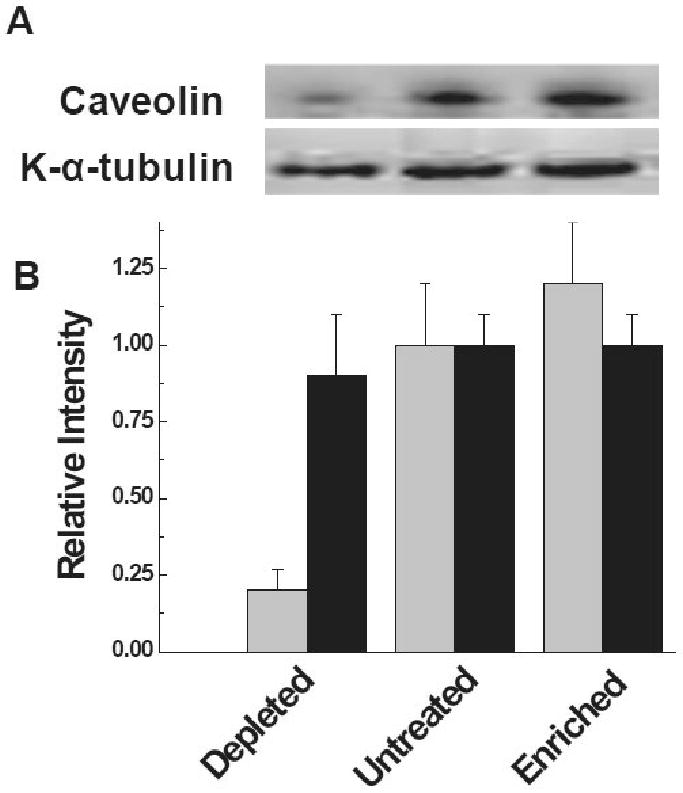
Figure 3.

Membrane localization of K-α-tubulin under changing membrane lipid raft domains. (A) Localization of K-α-tubulin and caveolin in the membrane fraction with and without treatment of βMCD. (B) Semi-quantitative morphometric analysis of the K-α-tubulin and caveolin; (light filled), represents caveolin and (dark filled), represents K-α-tubulin.
