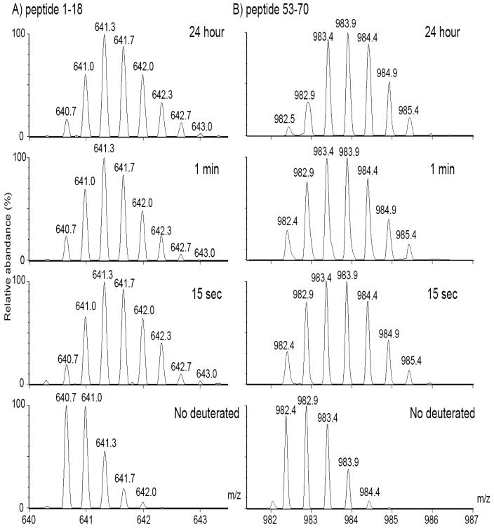
Fig. 2. Isotopic peak distributions of deuterated peptide masses (m/z) from WT β B2.
WT was incubated in D2O buffers for 0, 15 sec, 1 min, and 24 h and then digested with pepsin. A) The triply charged form of the N-terminal peptide, residues 1–18, shows a shift to higher mass at 15 sec with deuterium incorporation. No further increase in mass was detected with time. B) The doubly charged form of the interface peptide, residues 53–70, shows a more gradual shift in peak heights to a higher mass and therefore, a slower incorporation of deuterium.

