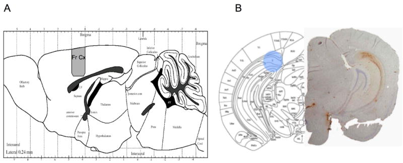
Figure 1.

A) Figure showing the region from which tissue was taken for both frontal cortical studies using the APP×PS1 and triple trasngene mice. B) Picture from a mouse brain atlas at the levels of the hippocampus (subiculum) showing the approximate region (blue) from which the tissue punches were taken. Shown on the right is the corresponding slice from a six month old mouse showing immunohistochemical stains for amyloid ß protein (Aβ1-42) in the hippocampus (shown in the figure on the right). The region from which the punch was taken is a region showing intense Aβ accumulation.
