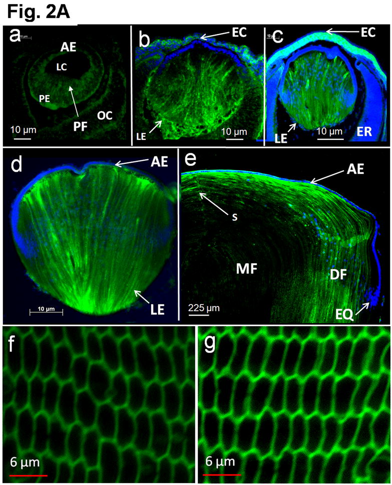
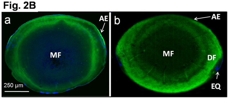
Fig. 2.


Transgene AQP1-EGFP expression in the developing mouse lens. A. Immunocytochemistry: Optical sections of TgAQP1+/+ eyes or lenses at: (a) E11.25; (b) E12.5; and (c) E15.5; (d) embryonic lens at E17.5 and (e) lens at postnatal day 8; (f) adult TgAQP1+/+ lens; (g) adult WT lens to show AQP0 expression (positive control for membrane localization). Anti-AQP1 antibody binding (green) to the differentiating and differentiated primary and secondary fiber cells (a-f). The antibody also bound to cornea, ciliary body and retina in embryonic eye. Anti-AQP0 antibody binding (green) to lens fiber cells (g). Selected lens sections were stained with DAPI (Blue) nuclear stain. AE – anterior epithelium; DF – differentiating fiber cells; EC – embryonic cornea; EQ – equatorial epithelial cells; ER – embryonic retina; LC – lens cavity; LE – lens; MF – matured fiber cells; OC – optic cup; PE – posterior epithelial cells; PF – primary embryonic lens fiber cells; S – fiber cell suture. B. Transgene AQP1-EGFP expression in adult mouse (3 months of age) lens. Immunocytochemistry: Optical sections of adult TgAQP1+/+ (a) and WT (b) mouse lenses to show patterns of transgene AQP1 and native AQP0 expression, respectively. In both cases, anti-AQP1 antibody bound (green) to the differentiating and differentiated secondary fiber cells and showed a similar pattern. There was an abrupt decrease in antibody binding at the transition from differentiating fibers to mature fibers, both in the TgAQP1+/+ and WT mice lenses.
