Fig. 6.


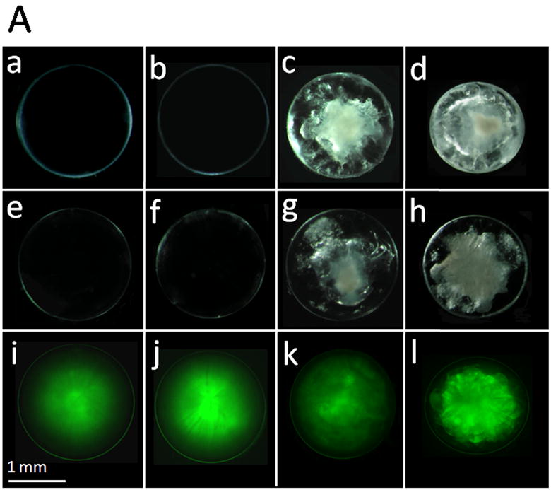
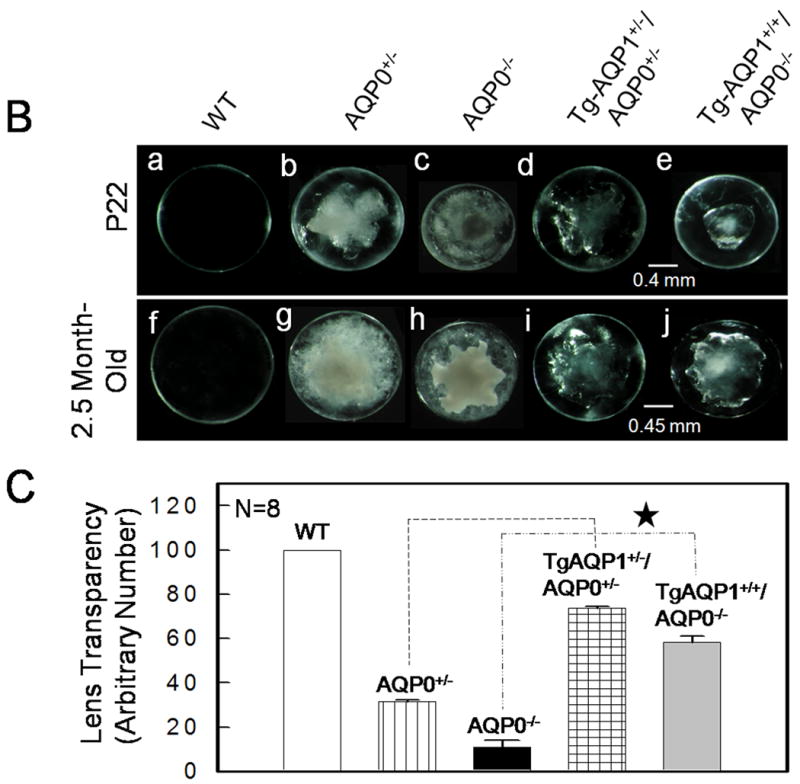
Lens transparency in relation to aquaporin expression. A. Lens transparency, cataract and AQP1-EGFP expression in 2-month old mice. (a,b), WT; (c), AQP0+/-; (d), AQP0-/-; (e,i), TgAQP1+/-; (f,j), TgAQP1+/+; (g,k), TgAQP1+/-/AQP0+/-; (h,l) TgAQP1+/+/AQP0-/-. (a-h), light microscopic images; (i-l), epifluorescent images of lenses expressing AQP1-EGFP chimeric protein. B. Relation between the age, and presence or absence of AQP0 and AQP1 in lens transparency and severity of cataract. Light microscopic images of lenses: (a,f), WT; (b,g), AQP0+/-; (c,h), AQP0-/-; (d,i) TgAQP1+/-/AQP0+/-; (e,j), TgAQP1+/+/AQP0-/-. C. Quantification of lens transparency at age P22 in the presence or absence of AQP0 and AQP1 in the lens fiber cells. N, number of lenses used to collect data. Star represents the degree of significance in comparison, P<0.0001.
