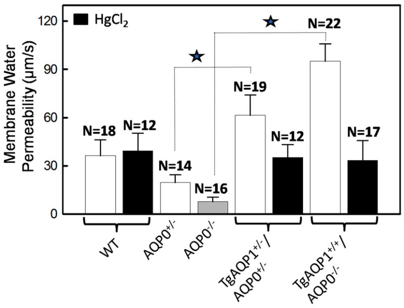
Fig. 7.

Lens membrane water permeability in the presence or absence of aquaporins. Fiber cell membrane water permeability of 4-month old WT, AQP0+/-, AQP0-/-, TgAQP1+/-/AQP0+/- and TgAQP1+/+/AQP0-/- mice. In some experiments, the fiber cell membrane vesicles were incubated with HgCl2 to inhibit AQP1. Each bar represents mean ± SD. N, number of membrane vesicles used per experiment. Star represents the degree of significance in comparison, P<0.0001.
