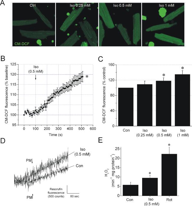
Figure 5. Isoflurane increases ROS production in cardiomyocytes and mitochondria.
(A) Representative images of CM-DCF fluorescence in cardiomyocytes. (B) Summary of signal traces of CM-DCF fluorescence intensity as indication of ROS production before and after addition of 0.5 mM isoflurane (Iso). (C) Summarized values of CM-DCF fluorescence intensity in control or after the treatment with various concentrations of Iso. (D) Representative traces of resorufin fluorescence intensity measurements in mitochondria with pyruvate and malate (PM) as substrate in the absence (Con) and presence of 0.5 mM Iso. (E) Summarized and calibrated rates of increase in resorufin fluorescence intensity in Con, Iso or rotenone (Rot, 1 μM)-treated mitochondria. Data are means ± SEM, N=6-10/group. *P<0.05 vs. Con.

