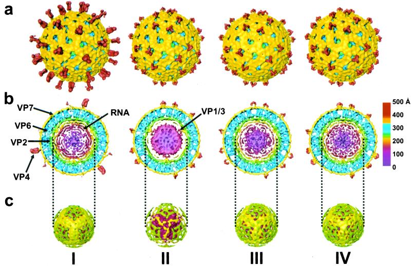
Figure 2.
Three-dimensional reconstructions of rotavirus under various chemical conditions. (I) TNC (pH 7.5). Arrows indicate various structural proteins and RNA. (II) 250 mM NH4OH (pH 11.5). Arrow indicates interaction between condensed RNA and VP1/VP3 complex. (III) TNC (pH 9.0) after NH4OH (pH 11.5). (IV) TNC (pH 7.5) after NH4OH (pH 11.5). The reconstruction is radially colored from the center to indicate the various protein layers according to the chart at the right. (a) Surface representations of the TLPs looking down the icosahedral 3-fold axis. The VP4 protein is red and VP7 is yellow. (b) Equatorial slice (≈47 Å thick) from the reconstruction. The VP6 protein is blue, VP2 is green, and the VP1/VP3 complex is red. The inner RNA layers are shown in colors ranging from yellow to maroon. (c) Surface representation at a radius of ≈233 Å showing the outer layer of RNA in yellow and the remnants of VP2 in green.

