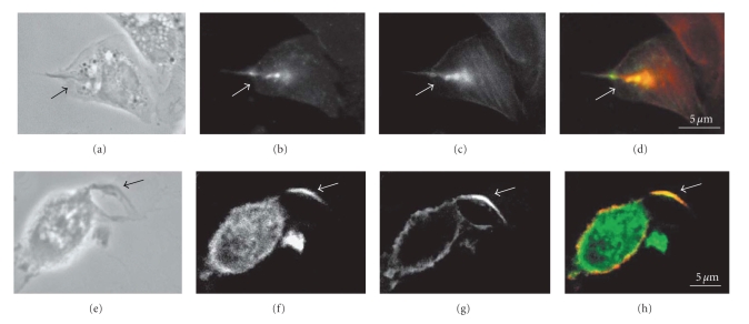
Figure 3.
Immunofluorescence microscopy colocalization (arrows) of phosphorylated proteins (b, g), detected using antiphosphotyrosine antibody, actin filaments (c), detected using phalloidin, and PI3 kinase (f) detected using anti-PI3 Kinase antibody during penetration of T. cruzi into macrophages. The overlay images (d, h) show the areas of colocalization demonstrating that phosphorilated proteins and microfilaments participate in internalization of T. cruzi trypomastigotes by macrophages. Bars = 5 μm (after [9]).

