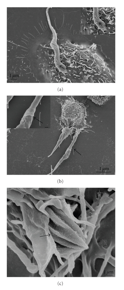
Figure 8.
Field emission scanning electron microscopy of the interaction between peritoneal macrophages treated with dynasore 60 μM (for 20 minutes) and allowed to interact with T. cruzi trypomastigotes (a), epimastigotes (b), and amastigotes (c). All parasite evolutive forms were partially recovered by the macrophage plasma membrane indicating that the blockage of GTPasic dynamin activity did not impair the pseudopod extension, impairing only the complete vacuole formation. The interaction time was enough to complete the parasite entry into control macrophages. Bars = 1 μm (after [29]).

