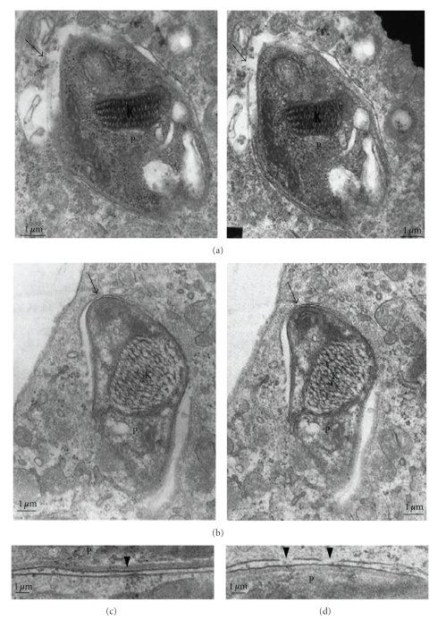
Figure 9.
Transmission electron microscopy (TEM) of thin sections of macrophages infected with trypomastigote forms of T. cruzi. Micrographs taken at different inclination angles of the section. Focal disruption of the membrane lining the vacuole is observed (arrows in (a) and (b)) and especially in (c) and (d); K = kinetoplast, P = parasite. Bars = 1 μm (after [52]).

