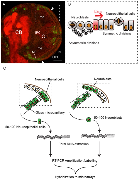
Fig. 1.
Symmetrically dividing neuroepithelial cells give rise to asymmetrically dividing neuroblasts. (A) Single confocal section of a third instar brain lobe showing the central brain (CB) and optic lobe (OL) stained for the neuroblast marker Dpn (green) and Dlg (red) to outline cells and the neuropile, respectively. The lateral half of each brain lobe comprises cells that form the developing optic lobe. Cells of the outer proliferation centre (OPC) give rise to the visual integration centres of the lamina and the outer medulla (me), whereas cells of the inner proliferation centre (IPC) generate the inner medulla, lobula and lobula plate. The OPC harbours two neural stem cell pools: lateral neuroepithelial cells and medial Dpn positive neuroblasts. Scale bar: 20 μm. (B) Optic lobe neuroepithelial cells divide symmetrically to expand the progenitor pool. The most medial neuroepithelial cells upregulate L'sc expression and are transformed into asymmetrically dividing neuroblasts. A wave of L'sc expression sweeps across the epithelium generating progressively more neuroblasts. (C) Genetically labelled neuroepithelial cells or neuroblasts were isolated from third instar larval brains using a glass microcapillary. Total RNA was extracted from 50-100 cells per sample and reverse transcribed, amplified by PCR and fluorescently labelled prior to hybridisation to a full genome microarray.

