Fig. 3.
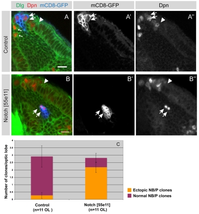
Notch mutant clones differentiate prematurely at aberrant positions. (A-A″) A single mCD8-GFP labelled control clone (blue) that contains several neuroblasts and progeny (A,A′). The clone originated in the neuroepithelium (to the right of the arrowhead) and has transformed into neuroblasts (arrows). Neuroblasts express the transcription factor Dpn (red in A,A″) and generate smaller progeny cells through asymmetric divisions (A,A′, arrows). Wild-type control neuroblasts are localised at the brain surface with the exception of the earliest born neuroblasts that migrate somewhat further into the cortex (A small arrows, A″). (B-B″) A representative mCD8-GFP labelled Notch loss-of-function clone is positioned aberrantly deep within the medulla cortex (B,B′ arrows). Two cells within this clone express Dpn (B,B″ arrows), which marks neuroblasts. Brains are counterstained for Dlg to outline cells. The arrowhead indicates the neuroepithelial to neuroblast transition zone. (C) Quantification of normal and ectopic neuroblast/progeny (NB/P) cell clones at 72 hours ALH. Virtually all control NB/P clones (91%, n=32) are located at the brain surface medial to the OPC neuroepithelium. By contrast, the great majority of Notch loss-of-function NB/progeny clones (77%, n=31) are localised ectopically within the medulla cortex. P<0.001 (unpaired t-test), error bars show s.e.m. Scale bars: 10 μm.

