Fig. 4.
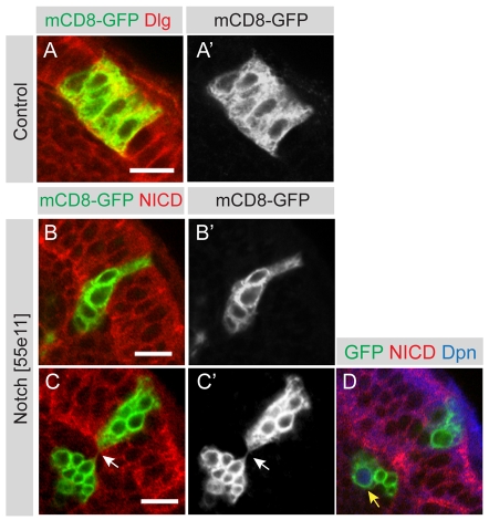
Notch mutant clones are extruded from the neuroepithelium. (A,A′) A single mCD8-GFP labelled control clone (green) was induced in the OPC, which is labelled by the septate junction protein Dlg (red). The clone contains several neuroepithelial cells generated through symmetric cell division. (B,B′) Shows a single mCD8-GFP labelled Notch loss-of-function clone (green) that is being extruded from the middle of the OPC neuroepithelium. Cells are stained with an antibody against the intracellular domain of Notch (red). (C,C′) A single mCD8-GFP labelled Notch loss-of-function clone (green) that has split in two. A group of cells has been extruded but is still attached by a thin membraneous connection (arrow) to clonally related cells that remain within the epithelium. (D) The same clone as in C, at a deeper section, shows a large cell with nuclear Dpn expression that has budded off smaller progeny cells. Scale bars: 10 μm.

