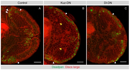
Fig. 7.
Disruption of Kuzbanian or Delta function leads to premature neuroblast formation. (A) A mid-third instar control brain is shown, stained for Dpn (green) to label neuroblasts and Dlg to outline cells (red). Dpn-positive medulla neuroblasts form at the medial edges of the OPC neuroepithelium (to the left of the arrowhead). (B) A mid-third instar Kuz-DN brain stained for Dpn (green) and Dlg (red). Disruption of Kuz function during the second larval instar transforms virtually all neuroepithelial cells into Dpn-positive neuroblasts. The IPC epithelium is disrupted and forms loop-like structures (yellow arrows), but exhibits no ectopic Dpn expression. Arrowheads indicate the putative neuroepithelial to neuroblast transition zone. (C) A late-third instar Dl-DN brain stained for Dpn (green) and Dlg (red). Disruption of Dl function during larval instars transforms neuroepithelial cells prematurely into Dpn-positive neuroblasts. Arrowheads indicate the putative neuroepithelial to neuroblast transition zone. Scale bars: 20 μm.

