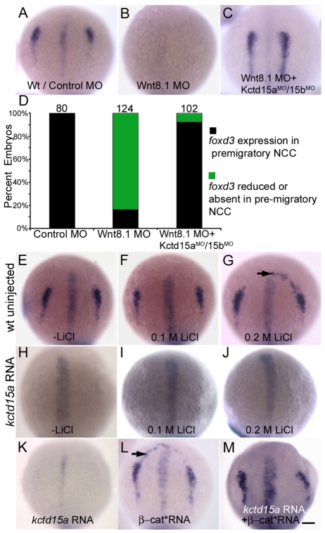
Fig. 3.
Kctd15 inhibits the output of canonical Wnt signaling. (A) foxd3 expression in control embryos. (B) Loss of foxd3 expression in wnt8.1 morphants. (C) Injection of Wnt8.1 MO together with Kctd15a/15b MOs rescued foxd3 expression. (D) Quantification of A-C; number of embryos (n) is given above bars. (E-J) foxd3 expression in embryos treated with LiCl (E-G). Arrow in G indicates expanded expression in 26/30 embryos. (H-J) LiCl did not rescue foxd3 expression in kctd15a mRNA-injected embryos (I, 40/40; J, 50/50; numbers indicate embryos with appearance as shown in the figure). (K-M) Inhibition of foxd3 by kctd15a (K) was rescued by β-cat* mRNA (M, 41/50 showing foxd3 expression in NC domain). β-cat* mRNA alone induced ectopic expression of foxd3 (L, arrow, 25/31). (A-C,E-M) 1-somite stage; dorsal views, anterior towards top. Scale bar: 100 μm.

