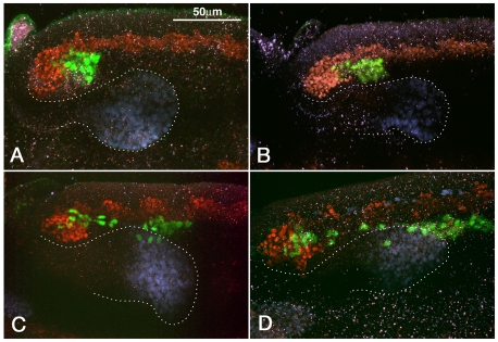
Fig. 3.
Early phase of migration of caudal visceral mesoderm cells. Sagittal views of posterior portion of elongated germ bands of HLH54Fb-lacZ Drosophila embryos stained with antibodies against β-gal (green; CVM), Twist (red; trunk mesoderm) and Hb9 (blue; midgut endoderm). (A) In stage 9 embryos, the posterior portion of the mesoderm bends around internally such that the CVM becomes positioned internally and slightly more anteriorly. (B) At stage 10, the CVM separates from the remaining mesoderm. (C) At late stage 10, individual cells leave the CVM clusters and start migrating anteriorly between posterior midgut (dotted outline) and trunk mesoderm. (D)By the end of stage 11, most CVM cells have surpassed the migrating endoderm anteriorly and are associated with trunk mesoderm.

