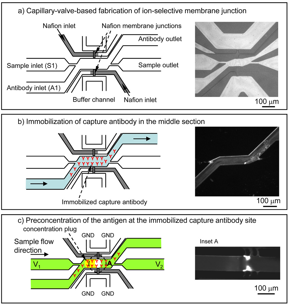
Fig. 2.
Schematic and fabrication of an integrated preconcentrator and surface-based immunoassay in microfluidic format: a) Filling of the Nafion resin into the junctions and subsequent curing, b) the side channel on the bottom (A1) was used to coat the surface of the sample channel with antibodies. After immobilization of capture antibody, sample was injected through the top side channel (S1). Since this channel was not surface-functionalized with antibody except for the area around the concentrator, sample depletion was minimized, as evidenced by the lack of binding in those regions. The insert shows a fluorescence image of R-Phycoerythrin molecules binding to only the coated region of the channel, c) preconcentration of RPE on the capture antibody site.

