
Figure 8. ELISA showing the expression of Aβ42 peptides in ear lysates.
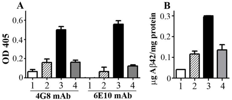
A Two Aβ42 monoclonal antibodies, 4G8 and 6E10, were used. Samples were analyzed in triplicates and shown are mean OD and SD. B To calculate the amount of peptide expression in the ears, an Aβ42 titration was used in the same ELISA. A threefold higher peptide concentration was demonstrated in ears transfected with Gal4/UAS-Aβ42 trimer compared to ears transfected with Gal4/UAS-Aβ42 monomer and CMV-Aβ42 trimer. White bars show results for ear lysates transfected with Gal4/UAS-Luc as negative control (1). Hatched and black bars show values for Gal4/UAS-Aβ42 monomer (2) and Gal4/UAS-Aβ42 trimer (3). Grey bars show expression levels for CMV-Aβ42 trimer (4).