Figure 4.

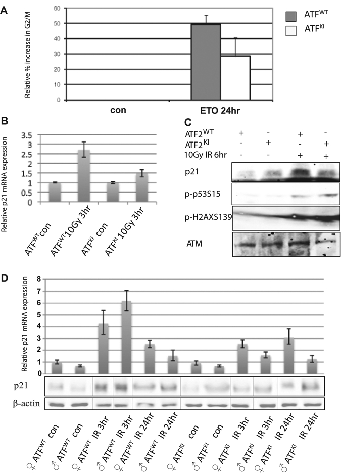
Expression of p21 is attenuated in ATF2KI cells and tissues subjected to genotoxic stress. (A) Murine embryonic fibroblasts (MEFs) prepared from wild-type (ATF2WT) and ATF2KI animals were subjected to treatment with etoposide (ETO), and cells were harvested 8 or 24 hours later for assessment of cell cycle distribution using fluorescence-activated cell sorting (FACS). Shown is the relative increase in the percentage of cells found to be in G2/M following ETO treatment. (B) Analysis of p21 transcripts was performed using qPCR analysis of cDNA prepared from RNA of the ATF2WT and ATF2KI MEFs at the indicated time points and doses of ionizing radiation (IR). (C) Western blot analysis of proteins prepared from ATF2WT or ATF2KI MEFs following IR (10Gy) or nonirradiated controls. Level of Ser15 on p53 and serine 139 on γH2AX (S139) served as controls for the cellular response to IR. Level of p21 proteins are shown (upper panel). (D) Real-time qPCR was performed on cDNA prepared from brain of ATF2WT or ATF2KI animals 3 and 24 hours after 8Gy IR. Representative experiment shown (out of 3 experiments, each using triplicates; error bars represent standard deviation). Western blots were performed with the same samples as the qPCR to determine the relative level of p21 protein.
