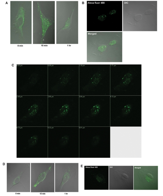
Figure 2.

EDC-Sulfo-NHS–Alexa Fluor 488 labeled IL-13-D2-NLS localizes to the nucleus of U-251 MG GBM cells. (A) U-251 MG cells were treated with 500 nM of the EDC-Sulfo-NHS–Alexa Fluor 488 labeled IL-13-D2-NLS and the subcellular localization monitored using Zeiss 510 LSM confocal microscope. (B) As in A, U-251 MG cell at 1 hour. DIC = differential image contrast for depicting the cell morphology. (C) Confocal Z-stack analysis of a U-251 MG cell treated with the labeled IL-13-D2-NLS protein at 1 hour. (D and E) U-251 MG cells were treated with 500 nM of the EDC-Sulfo-NHS–Alexa Fluor 488 labeled IL-13-D2 and the intracellular localization monitored using the Zeiss 510 LSM confocal microscope.
