Figure 1.
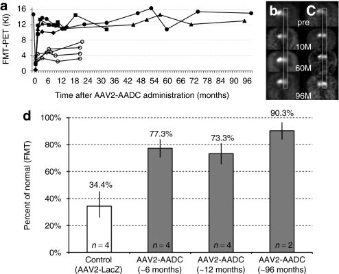
PET imaging of hAADC activity in hemi-lesioned monkey striatum. Data before 72 months are taken from Bankiewicz et al.4 (a) AADC activity (measured by Ki) over time. Open circles represent four control monkeys that received AAV2-LacZ after MPTP lesioning. Solid symbols indicated AAV2-hAADC-treated NHP; two NHP, Octopus (triangle) and Max (circles), remained in life out to 96 months. (b,c) PET images from AAV2-hAADC-treated animals, (b) Octopus and (c) Max. Images indicate coronal [18F]FMT PET images at baseline (pre) and at 10, 60, and 96 months after unilateral AAV2-hAADC administration to the right side of the brain. Both monkeys showed a stable and long-term increase in AADC activity. (d) Increase in FMT-PET signal after AAV2-AADC treatment over time. This restoration of signal was expressed as a percentage of the control/baseline FMT-PET signal (FMT uptake value in normal monkeys before MPTP lesioning). As there was no significant change in FMT uptake within the control or treated monkeys over time, we grouped animals into three time points (AAV2-AADC-treated monkeys; gray columns). The white column represents the mean FMT-PET signal from control monkeys, MPTP-lesioned monkeys and animals infused with AAV2-LacZ. FMT Ki values and earlier time points for the AAV2-AADC-treated monkeys were calculated from Bankiewicz et al.4 Standard deviations are shown for each group and the difference between the control and treated animals (at each time point) is statistically significant, P < 0.005. AADC, -amino acid decarboxylase; AAV, adeno-associated virus; FMT, 6-[18F]fluoro-meta-tyrosine; MPTP, 1-methyl-4-phenyl-1,2,3,6-tetrahydropyridine; NHP, nonhuman primate; PET, positron emission tomography.

