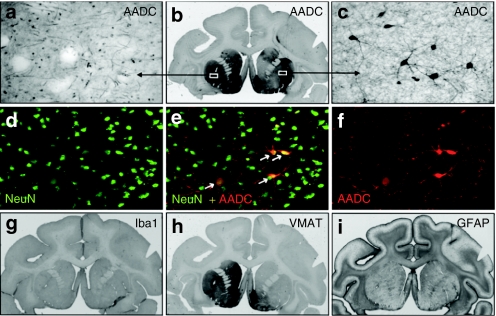
Figure 3.
Immunohistochemistry of the brain in hemi-lesioned monkeys 96 months after AAV2-AADC treatment. (a–c) AADC staining in one NHP (Max). The signal shows endogenous AADC in the unlesioned, left striatum, and almost complete restoration of the enzyme activity within the right striatum demonstrating the (b) long-term expression of AADC transgene. Endogenous AADC expression is characterized by (a) dense network of positive fibers whereas the restored lesioned side shows (c) transduced neuronal cell bodies along with the network of their processes. (d–f) Double immunofluorescent labeling of the AAV2-AADC-transduced hemisphere (Max) shows colocalization of markers AADC and NeuN (neuronal marker). The transduced neurons displayed a typical medium spinal morphology. (h) Staining against vesicular monoamine transporter (VMAT2), a marker for presynaptic neurons, shows a dramatic loss of dopaminergic terminals on the MPTP-lesioned, right hemisphere. The left side shows normal expression (compare to similar pattern of TH-staining—Figure 2a). (g) Staining for a microglia marker, Iba1, shows no inflammatory reaction on the AAV2-AADC treated side. Similarly, the lack of astrocytic activation was confirmed with (i) marker GFAP. AADC, -amino acid decarboxylase; AAV, adeno-associated virus; MPTP, 1-methyl-4-phenyl-1,2,3,6-tetrahydropyridine; NHP, nonhuman primate; TH, tyrosine hydroxylase.

