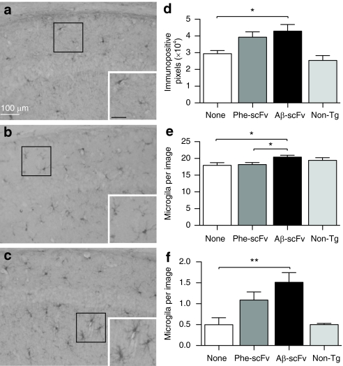
Figure 6.
AAV1 vector-mediated expression of the Aβ-scFv antibody increases number and activation of microglia. (a) Three-month-old 3xTg-AD mice received no injection, or (b) bilateral hippocampal injections of rAAV1-Phe-scFv or (c) rAAV1-Aβ-scFv. Mice were sacrificed at 12 months of age and processed for immunohistochemical analyses for IBA-1, which stains microglia. Nontransgenic animals were not included in statistical analyses, but used to demonstrate baseline levels of staining. Representative images are shown. (a) Bar =100 µm. Images were taken of the CA1 region of the hippocampus at ×20 magnification and numbers of (d) immunopositive pixels, (e) nonresting microglia, and (f) activated microglia were quantified. One-way ANOVA with Bonferroni's multiple comparisons post-tests were performed between treatment and control columns. One-way ANOVA (d) P = 0.0234; (e) P = 0.0186; (f) P = 0.0066. A capped line with an asterisk was used to indicate when two groups had statistically different means as determined by the post-test (*P < 0.05; **P < 0.01). 3xTg, triple transgenic; AAV1, adeno-associated virus type 1; AD, Alzheimer's disease; ANOVA, analysis of variance; Aβ, amyloid-beta; rAAV, recombinant AAV; scFv, single-chain variable fragment.

