
Figure 5.
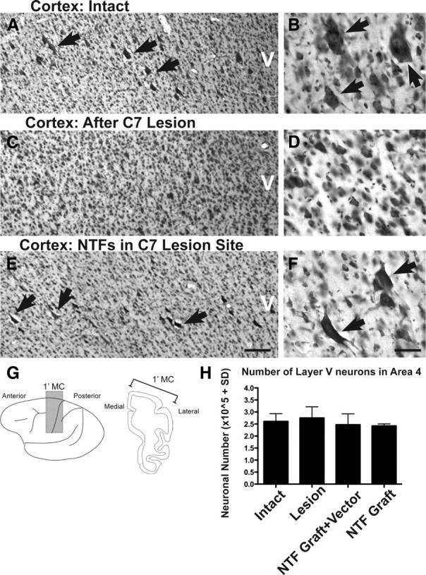
Cortical responses to injury and spinally administered neurotrophins. A, C, E, Representative thionin-stained coronal section from an intact monkey (A), lesion-only monkey (C), and lesion plus neurotrophin (NTF)-treated monkey (E). The arrows indicate large pyramidal neurons located within layer V. B, D, F, Higher magnification demonstrates a reduction in large layer V neurons after C7 hemisection that is prevented by growth factor administration to the spinal cord. G, H, Stereologic quantification of thionin-stained layer V total neuron number within the primary motor cortex (dark shading; G) indicates that total neuronal number in motor cortex is preserved after C7 lesions in all groups. Error bars indicate SEM. Scale bar: E (for A, C, E), 250 μm; F (for B, D, F), 25 μm.