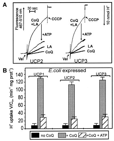
Figure 2.
Activation of H+ transport in UCP2 and UCP3 by CoQ10. H+ influx into phospholipid vesicles reconstituted with digitonin-treated IB-UCP2 and IB-UCP3 was recorded. (A) Recordings of H+ uptake in reconstituted UCP liposomes in the presence of CoQ10 (2 nmol) and absence of LA, with LA (125 μM) and without CoQ10, with CoQ10 and LA, and the inhibition with 20 μM ATP. (B) Evaluated H+ transport rates. H+ influx was measured as the change in external pH monitored by pyranine fluorescence at λex = 467 nm and λem = 510 nm in the presence of 125 μM LA. In all experiments (A and B) a 50-μl portion of vesicles containing 1.3 μg protein and 420 μg phospholipid was added to 0.5 mM Hepes buffer (pH 7.3) containing 1 μM pyranine, 0.5 mM EDTA, and 280 mM sucrose to a final volume of 330 μl at pH 6.8 and 10°C. H2SO4 was added in steps of 10 nmol H+ to adjust the pH to 6.8, and valinomycin (Val) was added to a concentration of 2.5 μM. Uncoupling by 1 μM carbonylcyanide m-chlorophenylhydrazone determined the capacity of H+ uptake of the vesicles. Results are presented as the initial H+ transport rate (V: μmol/min mg protein) divided by the capacity of the vesicles (C: μmol/ml).

