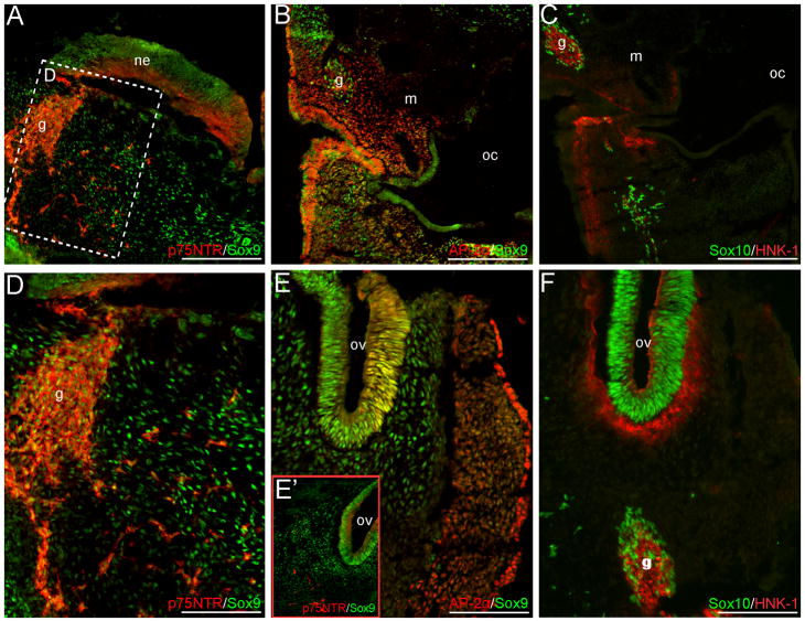
Fig. 3.
NCC markers are expressed in the mesenchyme or neural crest derivatives of cranial CS12 sections. Sox9 and AP-2α signal occur within the NCC-populated cranial mesenchyme or branchial arches (A–B, D–E’); panel in (D) is a higher magnification of the boxed region in (A). Additional Sox10+ and p75NTR+ cells are observed within this region (A, C–D, E’–F). NCC-derived cranial ganglia condensations or associated nerves express p75NTR, Sox9, Sox10, AP-2α and HNK-1 (A–D, F). Within the otic vesicles, Sox9, AP-2α, Sox10 and p75NTR signal occur (E–F). Axial level of sections is indicated by the green arrow in Fig. 1Q. Scale bars in (A–C) represent 200μm; those in (D–F) 100μm. g, ganglia condensation or associated nerve; m, mandibular/maxillay process; ne, neuroepithelium; oc, oral-pharyngeal cavity; ov, otic vesicle.

