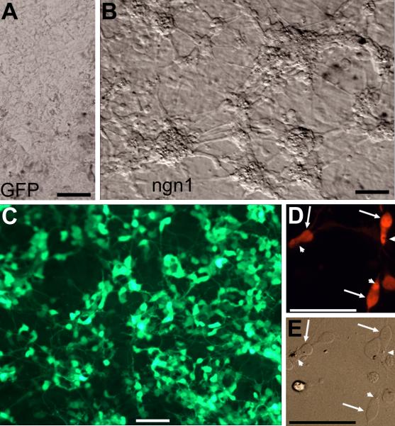
Fig. 1.
RPE cell cultures reprogrammed by ngn1. A,B: Bright-field views of a control culture infected with retrovirus RCAS-GFP (A) and a reprogrammed culture infected with RCAS-ngn1 (B). Red asterisks (*) in B mark cells clusters, which are absent in the control. C: Epi-fluorescence of visinin immunostaining of a ngn1-reprogrammed culture. D,E: Morphology of visinin+ cells viewed with bright-field (E) and epi-fluorescence (D). Arrows point to the cell body, and arrowheads point to a structural feature reminiscent of the lipid-droplet typically present in chick photoreceptors. Scale bars: 50 μm. A magenta-green copy is available as supplementary data.

