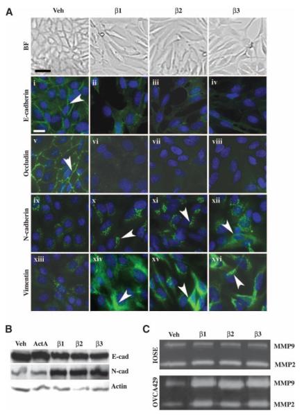
FIGURE 1.
TGF-β1, TGF-β2, and TGF-β3 mediate an EMT and MMP secretion in ovarian cancer cells. OVCA429 cells were serum-starved for 24 h and then treated with vehicle (Veh), 10 ng/mL TGF-β1, TGF-β2, TGF-β3, or activin A for 72 h. A. The TGF-β ligands stimulate an EMT in cancer cells. Top, brightfield images of cells showing that TGF-β initiates the loss of cell-cell junctions and the change from an epithelial to a fibroblastic morphology (bar, 20 μm); bottom, OVCA429 cells immunostained with anti–E-cadherin, anti-occludin, anti–N-cadherin, or anti-vimentin (FITC, white arrowheads) and counterstained with 4′,6-diamidino-2-phenylindole (blue) to show nuclei (bar, 50 μm). B. Anti–E-cadherin and anti–N-cadherin immunoblots of quiescent cells treated with vehicle, activin A, TGF-β1, TGF-β2, or TGF-β3. The same blot was stripped and reprobed with anti–β-actin as a loading control. C. TGF-β1, TGF-β2, and TGF-β3 trigger pro–MMP-2 and pro–MMP-9 secretion in cancer cells, but not in the normal cell line IOSE80. Conditioned media from quiescent OVCA429 and IOSE80 cells treated with vehicle, TGF-β1, TGF-β2, or TGF-β3 were subjected to gelatin zymography to analyze MMP secretion.

