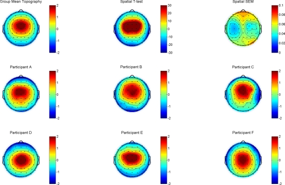
Figure 2.
Top row, left: the group average topography, scaled from −2 to +2 μV shows a clear maximum at FCz, extending to the neighboring electrodes FC, FC2, C1, Cz and C2. The spatial T-map (top row, middle) and the corresponding map of the standard error of the mean (SEM) illustrate the robustness of the selected topography in the sample. The middle and bottom rows (participants A–F) provide six randomly drawn single subject replications.

