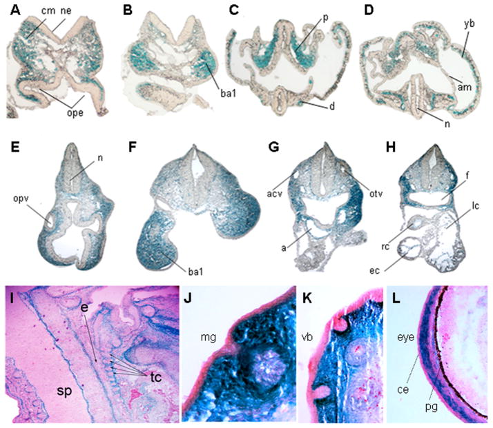
Figure 3. Tissue specific expression of the Inka1-LacZ allele.
A–H) Transverse sections of E8.0 (A–D) and E9.5 (E–H) embryos after whole-mount X-gal staining to detect expression of Inka1-LacZ. Plane of sections indicated by lines numbered in supplemental figure 1 (A–D) and Figure 2I(E–H). I–L) Sagittal sections of an E15.5 embryo stained with X-gal to show Inka1-LacZ expression and counterstained with nuclear fast red. a – aortic sac, acv – anterior cardinal vein, am – amnion, ba1– branchial arch 1, ce - conjunctival epithelium, cm – cephalic mesenchyme, d – dorsal aorta, e – esophagus, ec – endocardium, f – foregut, lc – left atrial chamber of heart, n – neural lumen, ne – neuroepithelium, mg – mammary gland, ope – optic evagination, opv – optic vesicle, otv – otic vesicle, p – paraxial mesoderm, pg – pigment layer of retina, rc – right atrial chamber of heart, sp – spinal cord, tc – throat cartilages, vb – vibrissae, yb – yolk sac blood island.

