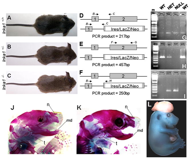
Figure 4. Inka1 function in mouse development.
A–C) Photographs of wild-type (A), Inka1 heterozygote (B), and Inka1 null (C) weanlings. D–F) Illustrations of RT-PCR reactions to examine splicing between exons 1 and 2 (D, PCR products shown in G), the deletion in exon 2 (E, PCR products shown in H), and splicing between exon 1 and exon 2 in the Inka1-LacZ allele (F, PCR products shown in I). Primers B, C, F, G, & H described in methods. J–K) Skeletal stains of PO wild-type (J) and Inka1−/− (K) heads. L) E15.5 Inka1-null mouse with exencephaly stained for LacZ expression. md – lower jaw, n – nasal bone, m – DNA marker, t – tympanic ring.

