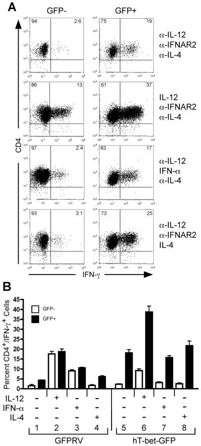
Figure 8. Ectopic T-bet expression promotes Th1 development independent of IL-12 or IFN-α in naïve human CD4+ T cells.
Purified naïve human CD4+ T cells were transduced with retrovirus constructs expressing GFP only (GFPRV) or with co-expression of human T-bet (hT-bet-GFP). During retroviral transduction, separate groups of cells were simultaneously activated in the presence or absence of cytokines or anti-cytokine antibodies as indicated in the figure. Cells were expanded on day 7 by restimulation on anti-CD3-coated plates. On day 14, resting cells were restimulated with PMA + ionomycin and analyzed for IFN-γ expression by intracellular cytokine staining. A, hT-bet-GFP-transduced cells were gated on live and either GFP negative (GFP-, left panels) or positive (GFP+, right panels) populations. The percentages of CD4+ and either IFN-γ- or IFN-γ+ populations are indicated within their respective quadrants. B, Triplicate cultures were analyzed for IFN-γ expression by intracellular cytokine staining. The percentage of CD4+/IFN-γ+ cells transduced with either the control GFPRV or hT-bet-GFP vectors are compared between the GFP- (open bars) and GFP+ (closed bars) populations. The error bars represent the standard deviation of the mean. These experiments were performed three times with similar results.

