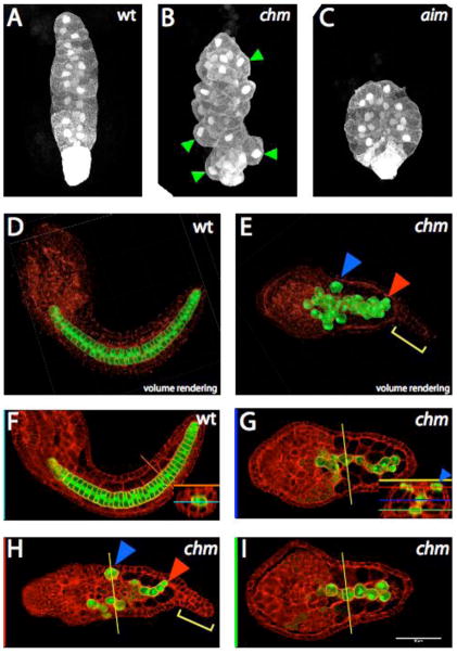
Fig. 1. Notochord morphology in chongmague.
All images show the notochord-specific bra:GFP transgene (white in A–C, green in D–I). Although not fused to any localization signal, the GFP is consistently brighter in the nucleus and cell cortex, and also brighter in the eight secondary lineage notochord cells at the posterior tip of the tail. Panels D–I also show the actin cytoskeleton labeled with phallacidin in red.
(A–C) Maximum intensity projections of confocal stacks through identically staged wildtype (A), chm/chm (B), and aim/aim (C) embryos. Anterior to the top. Green arrowheads in B show notochord cells at the edges of the notochord that have their long axes inappropriately oriented along the anteroposterior and not the mediolateral axis. (D, F) Mid-tailbud stage wildtype embryo. Anterior is to the left. (D) is a volume rendering of the entire confocal stack and (F) is a single slice at the level indicated by the blue line (inset). The inset shows a cross section along the orange line in the main panel. (E,G–I) chm/chm sibling. Anterior is to the left. (E) is a volume rendering and (G–I) are single slices at the levels indicated on the cross-section inset in G. The blue arrowheads mark an isolated notochord cell at the periphery of the tail. The red arrowhead marks a notochord cell interdigitated between two muscle cells. The yellow bracket marks the characteristic epidermal protrusion at the tip of the chm tail.

