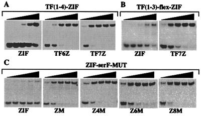
Figure 1.
A selection of DNA binding studies by gel-shift assay. (A) Shown are 5-fold dilutions of TF(1–4)-ZIF (from 5.5 nM–9 pM) against 20 pM ZIF binding site, 2 pM TF6Z, and 2 pM TF7Z. (B) Shown are 5-fold dilutions of TF(1–3)-flex-ZIF (from 5 nM–8 pM) against 20 pM ZIF and 2 pM TF7Z. (C) Shown are 5-fold dilutions of ZIF-serF-MUT (from 1 nM–1.6 pM) against 10 pM ZIF, 0.4 pM ZM, 0.4 pM Z4M, 0.4 pM Z6M, and 0.4 pM Z8M.

