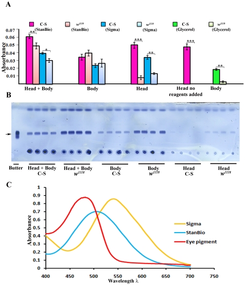
Figure 2. The coupled colorimetric triglyceride assay cannot accurately measure stored fat in adult Drosophila.
Whole w1118 (VDRC) flies contain more triglyceride than Canton S (C-S) flies, as shown by the stronger triglyceride bands (arrow) in (B) (head+body). Four replicates are shown. However, w1118 produces weaker signals in the colorimetric assay than C-S (A, head+body bars, when either the StanBio or Sigma kits are used). The bars represent an average of the values from four replicates. The TLC assay shows that, as expected, heads have much less fat than bodies. w1118 heads contain more fat than C-S heads (B, head). However, the colorimetric signal from C-S heads is much greater than that from w1118 heads. The same signal strength is observed when C-S heads are assayed without any added enzymes, showing that this signal is due to absorption by eye pigment. w1118 bodies produce a slightly stronger signal than C-S bodies, consistent with the TLC results; but the difference between C-S and w1118 bodies in the colorimetric assay is not statistically significant. We also measured glycerols in C-S and w1118 bodies, using the 'no lipase' reaction mix in the Sigma TR0100 kit. C-S bodies (bright green bars) contain much more glycerol than w1118 bodies (light green bars). Error bars are standard deviations of four different replicates for a given sample type. Asterisks denote T-test statistical significance: *; P<0.05–0,005, **; P<0.005–0.0005, ***; P<0.0005. (C) Absorption spectra of quinoneimines produced by the StanBio and Sigma kits, and of fly eye pigment.

