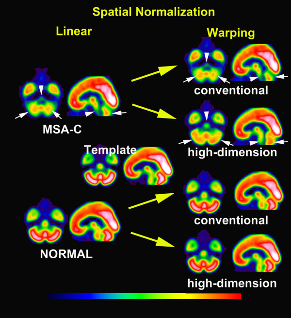
Figure 4.

Averaged images of spatially normalized with linear transformation and subsequent conventionally and high-dimensionally warped SPECT images in groups of MSA-C patients and normal controls. Linear spatial normalization showed insufficient transformation to a template in cerebellum (white arrows) and pons (white arrowheads) in MSA-C patients. Subsequent conventional warping could not perform a further transformation in these areas in this group. In contrast, high-dimension-warping could fully transform the cerebellum and pons to a template in MSA-C patients. Both conventionally and high-dimensionally warped SPECT images demonstrated decreased perfusion in the cerebellum and pons in MSA-C patients as compared to normal controls.
