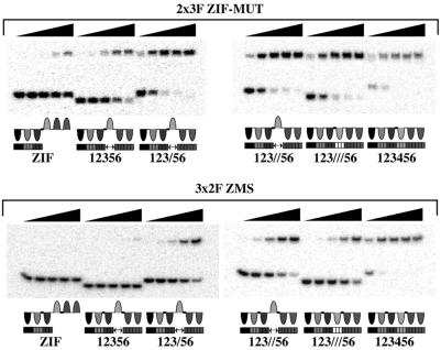
Figure 3.
A selection of DNA binding studies by gel-shift assay. The gels are designed to give a comparison between the binding affinities of the 2 × 3F ZM and 3 × 2F ZMS peptides and are not necessarily the gels used to quantify binding affinity. For example, the amount of 123456 binding site shifted by each peptide is limited by protein concentration rather than by Kd. (Top) Shown are 5-fold dilutions of 2 × 3F ZM (from 800 pM to 1.3 pM) against 2 pM binding sites. (Bottom) Shown are 5-fold dilutions of 3 × 2F ZMS (from 700 pM to 1.1 pM) against 2 pM binding sites. The proposed binding modes of the zinc finger peptides for each binding site is illustrated under each gel image. The lengths of oligonucleotides containing each binding site were as follows: 12356 and 123///56, 28 bp; ZIF, 123/56 and 123//56, 32 bp; and 123456, 34 bp.

