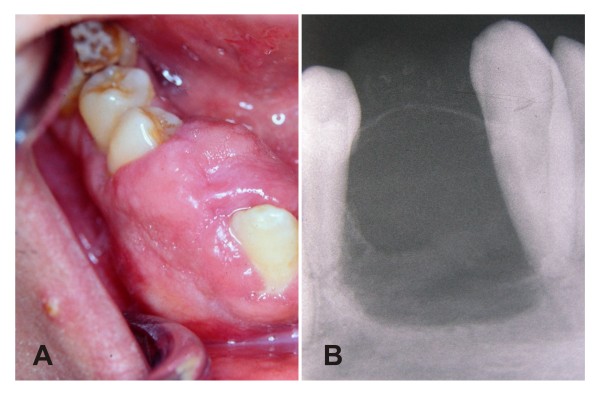
Figure 1.
A - Intra-oral view demonstrating gingival swelling in the alveolar ridge between canine and premolar teeth. B - Periapical radiography demonstrating a radiolucent osteolitic lesion with internal osseous septa, and points of calcifications on buccal surface. Besides, shows a thin radiopaque line around the superior aspect of the lesion and resorption of the lamina dura without radicular resorption.

