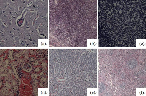
Figure 3.
Microscopic lesions in the coinfected group. (a) Perivascular cuffs in brain; (b) Suppurative pneumonia and purulent bronchitis; (c) Lymphocyte necrosis and disintegration in submaxillary lymph nodes; (d) Severe congestion of kidney and renal tubular necrosis and collapse; (e) Moderate sinusoidal congestion with a small number of lymphoid cells and macrophage infiltration in the lung; (f) Lymphoid cell necrosis and collapse around the ellipsoid arterioles and in the white pulp and red pulp of the spleen.

