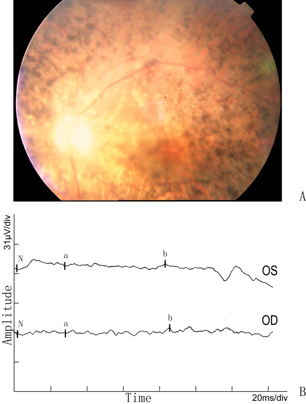
Figure 2.
Fundus photographs and ERG of the proband in the Chinese arRP family. The features of waxy-pale disc, arteriolar attenuation, and bone-spicule pigment deposit in the mid-peripheral retina are shown in A. The full-field ERG in the proband in B, either a-wave or b-wave has obvious reduced amplitude and prolonged response time in both eyes.

