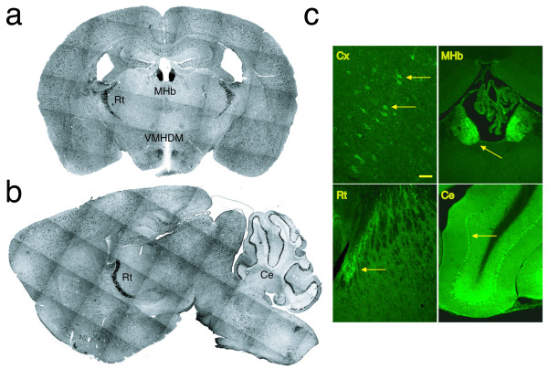
Figure 1.
KChIP1 expression in the adult mouse brain. a, b: In situ hybridization. Adult mouse brain sections were hybridized in situ with an anti-sense RNA probe synthesized using KChIP1 cDNA as the template. Coronal (a) and sagittal (b) views are shown. Hybridization signals are seen in a subset of neurons distributed in the cerebral cortex, hippocampus, and thalamus. High expression of KChIP1 was detected in the medial habenular nucleus (a, MHb), the reticular thalamic nucleus (a and b, Rt), the dorsomedial part of the ventromedial hypothalamic nucleus (a, VMHDM), and the Purkinje cell layer of the cerebellum (b, Ce). The montage was produced from photographs taken with a deconvolution microscope using a 5× objective and Slidebook software (Intelligent Imaging Innovations, Inc.). c: Immunohistochemical detection of KChIP1 in the brain. Adult mouse brain sections were immunostained with a polyclonal anti-KChIP1 antibody. Expression of KChIP1 was analyzed under confocal microscopy. The distribution of KChIP1 protein was similar to that of KChIP1 mRNA detected by in situ hybridization. KChIP1 was detected in a subpopulation of neurons in the cerebral cortex (Cx, yellow arrow); high levels of KChIP1 were observed in the medial habenular nucleus (MHb, yellow arrow), reticular thalamic nucleus (Rt, yellow arrow), and Purkinje cell layer of the cerebellum (Ce, yellow arrow). Bar = 50 μm in Cx and 100 μm in MHb, Rt, and Ce.

