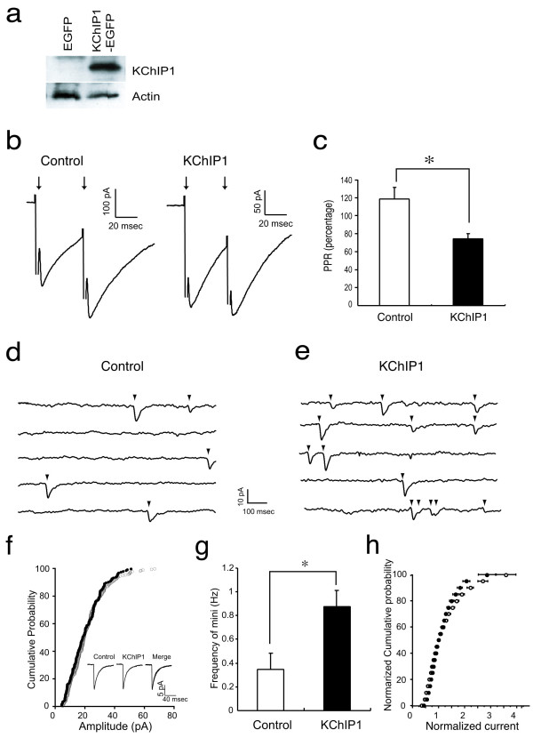
Figure 3.
Over-expression of KChIP1 facilitates GABA-mediated synaptic transmission in cultured hippocampal neurons. a: Expression of KChIP1-EGFP was detected by immunoblot analysis in neurons infected with KChIP1-EGFP (KChIP1) but not in neurons infected with EGFP alone. Detection of actin in the lysates with an anti-actin antibody is shown as a loading control. b: Representative recordings from EGFP+ (Control) and KChIP1-EGFP neurons (KChIP1) are shown. c: The paired pulse ratio (PPR) recorded in control cells was significantly larger than that seen in KChIP1-expressing cells (*P < 0.01). d, E: mIPSCs from autaptic inhibitory neurons. Representative mIPSC recordings are shown from EGFP+ (d) and KChIP1-EGFP+ neurons (e). f: Cumulative probability of amplitude and averaged mIPSCs (inset) from the same two cells as on f and e. g: The frequency of mIPSCs in cells expressing KChIP1 (0.88 ± 0.14 Hz) was more than twice that observed in control cells expressing EGFP alone (0.37 ± 0.13; *P < 0.05). h: There was no difference in the amplitude distribution of mIPSCs in these neurons (KChIP-EGFP: filled dots; EGFP alone: empty dots).

