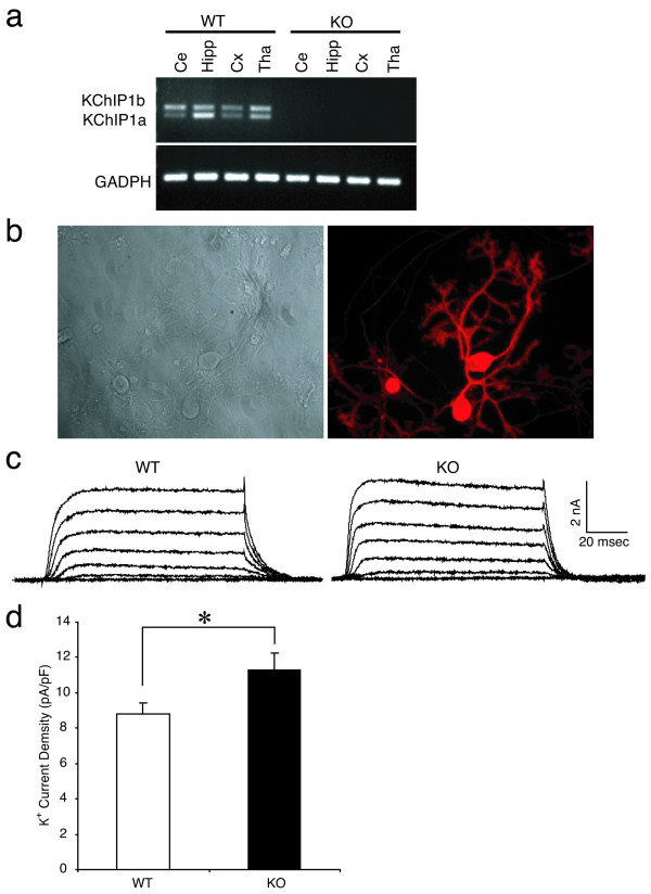
Figure 6.
Disruption of mouse KChIP1 gene potentiates potassium currents in cultured Purkinje neurons. a: Lack of KChIP1 mRNA in KChIP1-/- mouse brain tissue. KChIP1a and KChIP1b mRNAs in wild-type (WT) and KChIP1-/- (KO) mouse brain were analyzed by RT-PCR. The brain tissues examined include cerebellum (Ce), hippocampus (Hipp), cortex (Cx) and thalamus (Tha). Expression of GAPDH was monitored as a positive control. b: Primary cultures of Purkinje cells from KChIP1-/- mouse cerebella. A phase contrast image (left panel) and an immunofluorescence image of Purkinje cells stained by with anti-calbindin antibody (right panel) are illustrated. c, d: Potentiation of potassium currents in KChIP1-/- Purkinje cells. Representative recordings from wild-type (WT) and KChIP1-/- (KO) Purkinje cells (c). Potassium current density in WT was significantly smaller than that seen in KO neurons (d, *P < 0.05). Whole-cell potassium currents were evoked by a voltage step to -15 mV from a holding potential of -75 mV. Steady-state amplitudes were normalized to cell capacitance.

