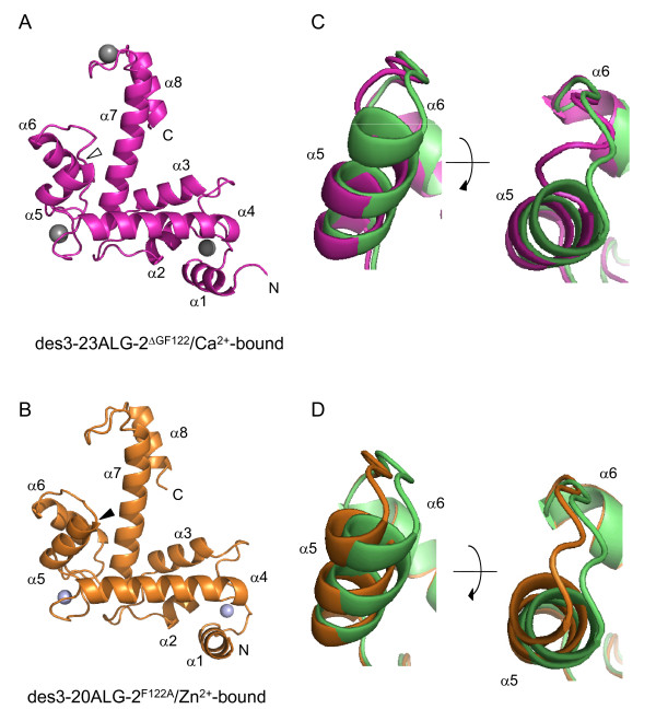
Figure 1.
Structures of ALG-2ΔGF122 and ALG-2F122A. Structures of (A) Ca2+-bound des3-23ALG-2ΔGF122 (PDB ID 3AAJ) and (B) Zn2+-bound des3-20ALG-2F122A (PDB ID 3AAK) are shown in magenta and orange, respectively, in ribbon representations, and EF-hand-coordinated calcium and zinc ions are shown in gray and light cyan spheres. The deleted site (Gly121Phe122) and substituted site (F122A) are marked by an open arrowhead and closed arrowhead in (A) and (B), respectively. (C, D) The ALG-2 structures are compared with the previously resolved structures of wild-type ALG-2 of Ca2+-bound form (PDB ID 2ZN9) and Zn2+-bound form (PDB ID 2ZN8) by aligning at α-helix 4 (α4), and close-up views of segments from α5 to α6 are shown in ribbon representations. (C) The structures of Ca2+-bound des3-20ALG-2 and des3-23ALG-2ΔGF122 are shown in green and magenta, respectively. The view in the left panel is rotated approximately 90° to view from the top as shown in the right panel. (D) The structures of Zn2+-bound ALG-2 (green) and des3-20ALG-2F122A (orange) are presented similarly to (C).

