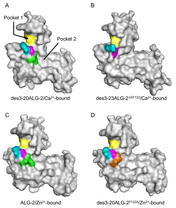
Figure 3.
Loss of a wall surrounding hydrophobic pockets in ALG-2ΔGF122. Surface structures of (A) Ca2+-bound des3-20ALG-2, (B) Ca2+-bound des3-23ALG-2ΔGF122, (C) Zn2+-bound ALG-2, and (D) Zn2+-bound des3-20ALG-2F122A are presented in gray except for indicated residues of Gly121Phe122 (green), G123 (or G121 in des3-23ALG-2ΔGF122) (magenta), A122 in the F122A mutant (orange), R125 (or R123 in des3-23ALG-2ΔGF122) (cyan), Y180 (or Y178 of des3-23ALG-2ΔGF122) from a dimerized counterpart molecule (yellow).

