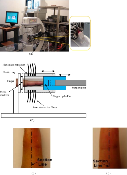
Figure 1.
(a) Photograph of the integrated hybrid x-ray∕DOT system. The inset (right part) is a close-view photograph of the finger∕fiber optics∕x-ray interface. (b) Schematic of the interface. Note that both the Plexiglass container and finger tip holder can be translated horizontally for separate DOT and x-ray data acquisition. (c) The sagittal plane of a 3D DIP finger joint. (d) The coronal plane of a 3D DIP finger joint.

