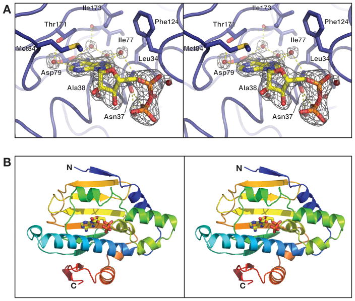
Figure 1. Overall structure and nucleotide binding by Hsp90N.
(A) Close-up of the Hsp90N nucleotide-binding site bound to ADP. Amino acid residues surrounding the active site are shown in stick view, and water molecules involved in hydrogen bonding are shown as spheres. Fo-Fc simulated-annealing omit electron density is shown for ADP and surrounding water molecules, contoured at 6.0 σ. (B) Stereo view of monomer A of Hsp90N, bound to ADP. The protein is colored in a rainbow scheme, with the N-terminus blue and the C-terminus red. Bound ADP is shown in stick view.

